Canberra Equine Hospital offers an array of up-to-date, modern diagnostic imaging modalities to assist in obtaining information and diagnose the problem.
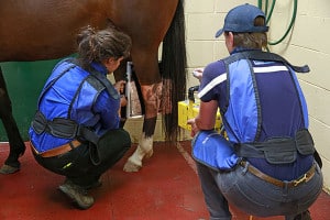
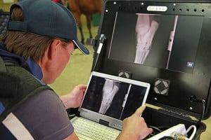
 Radiology: We utilise two advanced, portable X-RAY units to obtain instant, high-quality digital images and aid an accurate diagnosis. X-RAYs can be performed in hospital or on farm. Images can be emailed to you or your regular veterinarian if a case is referred. Thorough reports and recommendations are provided with X-RAY evaluations.
 Prepurchase Radiology: A full series of radiographs or few images focused on potential problem areas can be taken as part of a prepurchase examination.
Yearling Radiology: It is common practice for Thoroughbred yearlings to have thorough pre-sale radiographic examinations. 34 views for Australia, and 38 for Hong Kong, are obtained of feet, fetlocks, knees, hocks and stifles. Accurate, high quality images are provided to the repository veterinarians, assisting with a fast interpretation, as well as improving the saleability of yearlings.
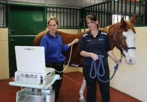
 Diagnostic Ultrasound: A non-invasive way to diagnose soft tissue injuries, such as tendon or ligament tears, haematomas, abscesses and tumours. Also useful to image the chest (e.g. pneumonia, foals with “Rattles”), and abdomen when making an assessment of a horse with colic. Echocardiography, or evaluations of the heart, are also performed with ultrasound.
Video Endoscopy: A long tube with a little video camera and light on the end is passed into the airway, (pharynx, larynx, windpipe and lungs), or the oesophagus and stomach. This allows us to view the structure and function of a horse’s airway and stomach. A horse experiencing poor performance may have a respiratory dysfunction such as making noise from a collapsing laryngeal cartilage, (a “Roarer”), be bleeding during high intensity work or have a structural abnormality. The stomach and oesophagus are often assessed for presence of ulceration. We have the ability to use endoscopes in the field or at our hospital.
